Mammogram vs Breast MRI for Cancer Screening What You Need to Know

Breast cancer screening is not one single test. Mammograms and breast MRIs can both find cancer, but they work in very different ways, feel different during the appointment, and serve different roles in care.
For many people, mammography is the main screening tool. Breast MRI is usually reserved for people with a higher risk of breast cancer or for certain follow-up situations. Knowing how each test works can make screening decisions feel more understandable and less abstract.
This article is for general education only. It does not replace medical advice from your clinician who knows personal risk factors, prior imaging, family history, and overall health.

How mammograms work
A mammogram uses low-dose X-rays to create images of breast tissue. The goal is to find signs of cancer before a lump can be felt or symptoms appear.
During the exam, a technologist places one breast at a time between two plates. The machine compresses the breast for a brief period while it takes images from different angles. Compression can be uncomfortable, but it helps spread the tissue out so small changes are easier to see. It also reduces motion and allows the machine to use a lower radiation dose.
Most screening centers now use digital mammography. Most also use 3D mammography, also called breast tomosynthesis. With 3D mammography, the machine takes images from multiple angles and a computer builds thin image slices. This can help radiologists see through overlapping tissue, especially in people with dense breasts.
A screening mammogram is used when there are no symptoms. A diagnostic mammogram is used when there is a symptom, such as a lump, nipple discharge, skin change, or an abnormal screening result. Diagnostic imaging may include extra mammogram views and often a breast ultrasound.
How breast MRI works
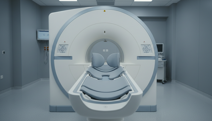
A breast MRI uses a strong magnet and radio waves, not X-rays. It creates detailed images of breast tissue and blood flow patterns. Most breast MRI exams use an injected contrast agent, usually gadolinium-based contrast, to help highlight areas that may need closer review.
During the exam, the person lies face down on a padded table with the breasts positioned in openings in a special breast coil. The table slides into the MRI scanner. The test is painless, but the scanner can be loud, and the space can feel narrow. Ear protection is typically provided. The exam usually takes longer than a mammogram.
MRI is very sensitive. That means it can detect many cancers, including some that may not show clearly on mammography. The same sensitivity also means MRI can find areas that look suspicious but are not cancer. Those findings can lead to additional imaging or biopsy.
Breast MRI is not usually used alone for routine screening in average-risk people. In many high-risk screening plans, MRI is used in addition to mammography, not as a replacement.
What the appointment feels like
The two tests differ not only in technology, but also in the practical experience.
A mammogram is usually short. The imaging itself takes only a few minutes, though the full appointment takes longer because of check-in, changing clothes, and positioning. The breast compression lasts only seconds for each image. Some people feel pressure or pain, especially if the breasts are tender, but the discomfort passes quickly.
A breast MRI takes more time. The person must stay still while the scanner captures image sequences. The machine makes loud tapping or thumping sounds. Some people find the face-down position uncomfortable. People with claustrophobia may need extra planning, such as discussing relaxation options with the imaging center before the appointment.
MRI also requires safety screening. Because the scanner uses a powerful magnet, the imaging team checks for implanted devices, metal fragments, certain surgical clips, and other items that may not be safe in an MRI environment. When contrast is used, the care team also considers kidney function, prior contrast reactions, pregnancy status, and other medical factors.
The main strengths of mammograms
Mammography has been studied for decades and remains the standard screening test for most people at average risk. Its biggest strength is that it can find early signs of breast cancer, including tiny calcium deposits called calcifications. Some calcifications can be linked to ductal carcinoma in situ, or DCIS, which may appear on a mammogram before it causes symptoms.
Mammograms are also widely available across the United States. Many hospitals, breast centers, and mobile screening programs offer them. Appointments are usually shorter and less expensive than MRI exams. Insurance coverage for screening mammography is also well established for eligible age groups, although details vary by plan.
Another advantage is consistency. Because mammography is so commonly used, radiologists can compare current images with prior mammograms. This helps them see whether an area is stable or changing over time. Stability can reduce unnecessary follow-up, while change can prompt closer evaluation.
The limitations matter too. Mammograms use a small amount of ionizing radiation. For most people, the benefit of regular screening outweighs this low exposure, but it remains part of the risk-benefit discussion.
Mammography can also be less sensitive in dense breast tissue. Dense tissue and tumors can both appear white on a mammogram, which can make cancers harder to see. Dense breasts are common, and breast density is now routinely reported to patients in the United States. Having dense breasts does not automatically mean MRI is needed, but it may lead to a discussion about supplemental screening based on overall risk.
False positives are another drawback. A mammogram may find an area that looks concerning but turns out not to be cancer. This can lead to extra imaging, short-term follow-up, or biopsy. False negatives can also occur, meaning a cancer is present but not detected on the mammogram.
The main strengths of breast MRI
Breast MRI is highly sensitive. It can find cancers that may be hidden on mammography or ultrasound, especially in higher-risk patients and in dense breast tissue. For people with a strong inherited risk, that added sensitivity can be important.
MRI also does not use ionizing radiation. This makes it useful when repeated imaging is needed in certain high-risk groups, especially when screening starts at a younger age.
Another strength is the amount of detail it provides. MRI can show how an area takes up and releases contrast over time. Radiologists use these enhancement patterns, along with shape and margins, to decide whether an area looks suspicious. MRI can also help evaluate the extent of known breast cancer in selected cases, though that is diagnostic use rather than routine screening.
MRI has clear limitations. It is more expensive and less widely available than mammography. The appointment is longer. Some people cannot have MRI because of implanted devices or metal safety concerns. Others may not be able to receive contrast.
MRI is also more likely to detect findings that are not cancer. Because it is so sensitive, it may lead to extra tests and biopsies. For people at average risk, this can create more harm than benefit. That is one reason breast MRI is not recommended as a routine screening test for everyone.

When mammography is usually recommended
For people at average risk, mammography is the usual screening test. In the United States, many guidelines now recommend starting regular screening mammograms at age 40, although the exact schedule can vary by medical organization and personal preference. Some recommend yearly screening, while others recommend every two years.
Average risk generally means no known high-risk gene mutation, no strong family history suggesting inherited risk, no prior chest radiation at a young age, and no personal history of high-risk breast lesions or breast cancer. Our providers can help define risk more precisely.
Mammography may also be recommended earlier or more often when risk is above average but not high enough to require MRI. Personal history matters. So do family history, breast density, prior biopsy results, and risk model estimates.
A screening mammogram is not the right test for every breast concern. If there is a new lump, nipple discharge, breast pain in one area, skin dimpling, or nipple change, diagnostic evaluation is needed. That may include diagnostic mammography, ultrasound, or other imaging depending on age and symptoms.
When breast MRI is usually recommended
Breast MRI is most often recommended for people at high risk of breast cancer. This can include people with a known BRCA1 or BRCA2 mutation or another high-risk inherited mutation. It can also include people with a very strong family history, prior radiation to the chest at a young age, or a calculated lifetime breast cancer risk above a threshold used by many guidelines.
High-risk screening often starts earlier than average-risk screening. In many cases, breast MRI begins in young adulthood, and mammography is added later. The plan depends on the specific risk factor and the clinician’s judgment.
MRI may also be considered for some people with dense breasts, especially when dense tissue combines with other risk factors. It is not automatically recommended for every person with dense breasts because risk varies widely.
Some people with a personal history of breast cancer may have MRI as part of follow-up, especially if they were diagnosed young, have dense breasts, or have other risk factors. MRI can also be used before surgery in selected newly diagnosed patients to evaluate the extent of disease, but that is different from screening.
The key point is that MRI is a targeted tool. It can be very helpful for the right person, but it is not the default screening test for the general population.
How the two tests can work together
The phrase Mammogram vs Breast MRI for Cancer Screening What You Need to Know may sound like an either-or choice, but many screening plans are more nuanced. For average-risk screening, mammography usually stands on its own. For high-risk screening, mammography and MRI often work as partners.
Mammogram | Breast MRI |
Uses low-dose X-rays to image breast tissue. | Uses magnets, radio waves, and usually contrast. |
Best established for average-risk population screening. | Most often used for high-risk supplemental screening. |
Can show calcifications that may be early signs of disease. | Can find some cancers hidden by dense tissue or overlapping structures. |
Shorter, more available, and usually less costly. | Longer, more sensitive, and more likely to lead to extra follow-up. |
One test can detect findings the other may miss. Mammography is especially useful for calcifications. MRI is especially useful for certain invasive cancers that enhance with contrast. This is why high-risk screening plans may include both rather than replacing one with the other.
What to ask before choosing a screening plan
The best screening plan starts with risk. Age alone is not the full picture. Family history on both sides of the family matters. So do genetic test results, prior breast biopsies, breast density, previous cancer treatment, and hormone-related factors.
This is why our providers at The Breast and Wellness Collective ensure a complete evaluation of your specific situation and circumstances to determine whether your risk is average, increased, or high. We put together a surveillance plan specific to you to determine when to start screening, how often to screen, and whether supplemental imaging makes sense.
Screening decisions can also change over time. A new family diagnosis, genetic test result, biopsy finding, or change in health status may shift the plan. At The Breast and Wellness Collective we continually review, monitor and update your specific care plan based on changing details.

Frequently Asked Questions
Is a breast MRI better than a mammogram?
Not for everyone. MRI is more sensitive, but that does not always make it the better screening test. For average-risk people, mammography remains the standard because it has strong evidence, broad availability, and a better balance of benefits and harms. MRI is most useful when breast cancer risk is high or when a clinician has a specific reason to recommend it.
Can breast MRI replace mammography?
Usually no. In high-risk screening, MRI is often added to mammography rather than used instead of it. Mammograms can show calcifications that MRI may not detect as clearly. The two tests provide different types of information.
Is mammogram radiation dangerous?
Mammograms use a low dose of radiation. For most eligible people, the benefit of finding breast cancer early outweighs the small radiation risk. People who have concerns about radiation exposure should discuss their personal risk and screening schedule with a clinician.
What if I have dense breasts?
Dense breasts can make mammograms harder to read and are also linked with a higher risk of breast cancer. Many people with dense breasts still use mammography as their main screening test. Supplemental imaging, such as MRI or ultrasound, may be considered based on overall risk, not density alone.
Why would an MRI lead to more follow-up tests?
MRI can detect very small or subtle areas of enhancement. Some of these areas are cancer, but many are benign. If an area looks uncertain, the radiologist may recommend more imaging, short-term follow-up, or a biopsy to make sure.
The key takeaway
Mammograms and breast MRIs are both valuable, but they are not interchangeable. Mammography is the foundation of screening for most average-risk people. Breast MRI plays a major role for people at high risk and in selected situations where more detail is needed.
The most useful next step is to understand personal risk. With the information we obtain about you and your history, our providers recommend a screening plan that fits the person, not just the test. Regular screening, done at the right time with the right method, gives breast cancer the best chance of being found early.
Disclaimer: This article provides general information and is not a substitute for professional medical advice. Always consult your healthcare provider for personalized care.


Comments